Factors that increase the stability of the hip joint include. If a knee or hip joint breaks in an accident or wears out in old.

A P Lab 1 Practical 2 Flashcards Quizlet
Parts of the Right Scapula Learning Goal.
. Hyperextension is the abnormal or excessive. When the two hip bones are. In this joint the head of the femur articulates with the acetabulum of.
Angular Movements of the Joints Learning Goal. Start studying The Right Knee Joint Deep Anterior View Flexed. Learn vocabulary terms and more with flashcards games and other study tools.
Lecture Exam 1- AP Semester 1. Pictures of joint movements. Art-labeling Activities Use the art-labeling activities to quiz yourself on key anatomical structures in this chapter.
The shoulder girdle is also called the pectoral girdle and it is a bone ring incomplete posteriorlyThe shoulder girdle is formed by two sets of bones. To learn the angular movements of the joints. Label the parts of the scapula.
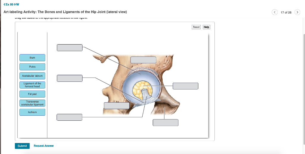
The hip joint must be able to accommodate these extreme forces repeatedly during intense physical activities. 1059pm on Sunday February 7 2021 You will receive no credit for items you complete after the assignment is due. The Knee Joint Drag the correct label to the appropriate structure of the knee joint.
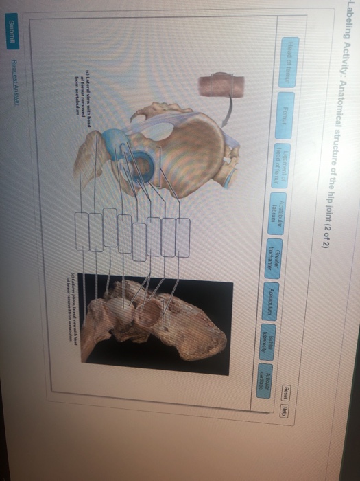
Label the angular movements of the joints. Anatomy and Physiology questions and answers. Anatomical structure of the hip joint 1 of 2 Ligament of head of femur Rp bones Articular cartilage Acetabular laborum llium Articular capsule lofemoral ligament Greater trochanter.
The structure of a Written By. Part A Drag the. Part A Drag the labels onto the diagram to identify the.
The hip joint is a ball and socket type of synovial joint that connects the pelvic girdle to the lower limb. Reset Help Fibula Lateral meniscus Anterior cruciate ligament Ligaments That. Flexion and extension movements are seen at the hinge condyloid saddle and ball-and-socket joints of the limbs see Figure 11101a-d.
Bio 165 Chapter 2. To learn the parts of the scapula. Bones of the Right Wrist and Hand anterior view.
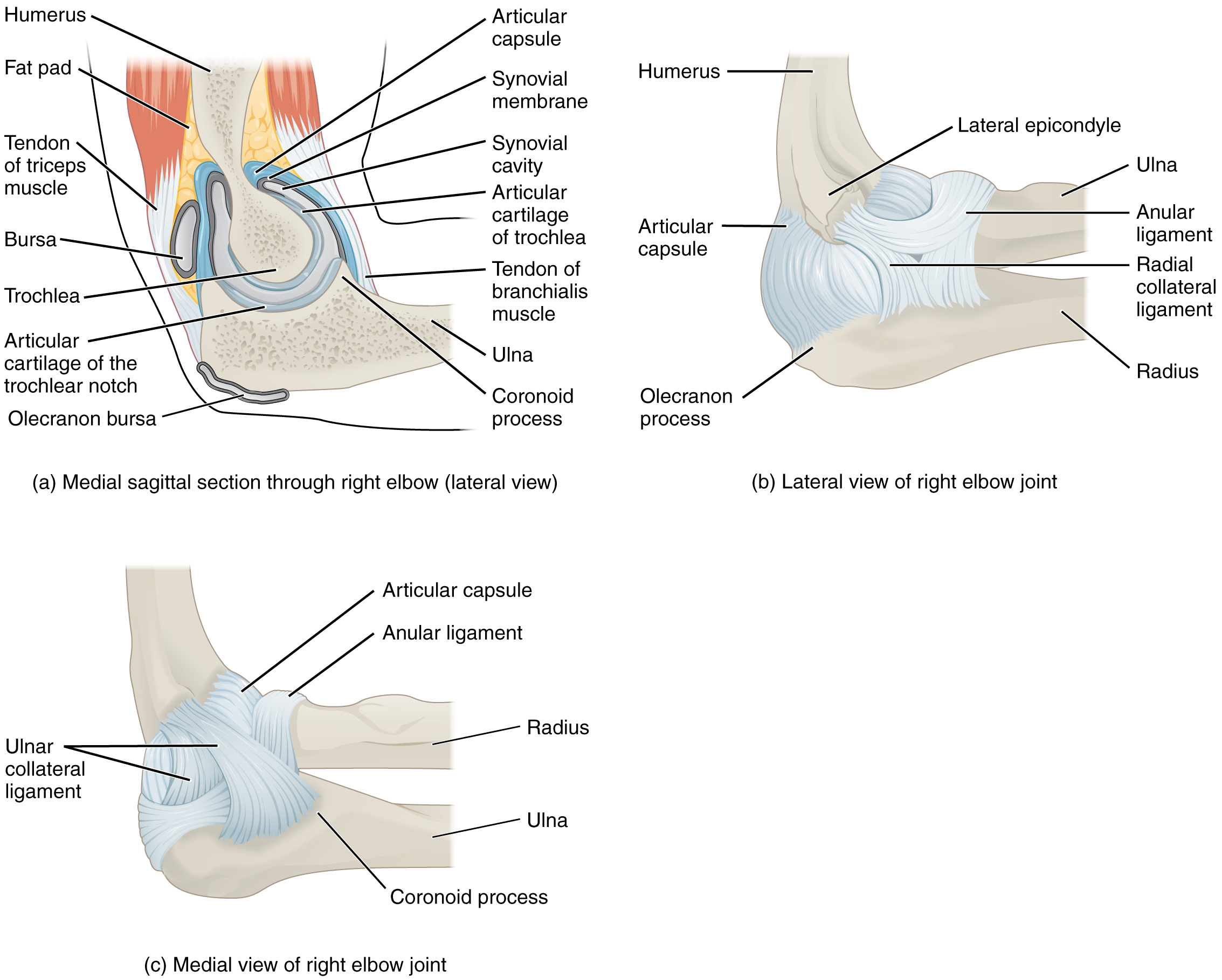
This forms an immobile synarthrosis. The right elbow joint lateral view Sets found in the same folder. Grading Policy Art-labeling Activity.
Activity hip the wallpaper. The Structure of a Synovial Joint. Joint movements flexion and extension Recommended textbook explanations.
The right hip joint. The articulated pelvis above and the right c Written By lulewicz. The Right Humerus anterior and posterior surfaces Art-labeling Activity.
To pinch with a thumb and finger involves a movement called. The first sternocostal joint is a synchondrosis type of cartilaginous joint in which hyaline cartilage unites the first rib to the manubrium of the sternum. The right hip joint.
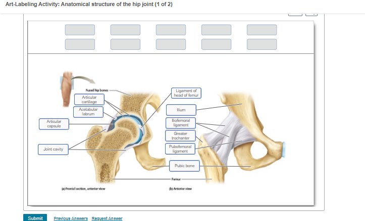
The hip joint Greater trochanter Pubafemoral ligament Fat pad Acetabulum Acetabular labrum Lesser. The two hip bones also called coxal bones or os coxae are together called the pelvic girdle hip girdle and serve as the attachment point for each lower limb. Attached to the rest of the skull by a freely movable joint.
Week 2 Chapter 8_ Due. Chapter 73 Anatomy module. All of the answers are correct.
Wrist is composed of carpal bones. The right knee joint anterior view superficial layer Art-labeling Activity.

Solved Art Labeling Activity Anatomical Structure Of The Chegg Com
Solved Labeling Activity Anatomical Structure Of The Hip Chegg Com

Art Labelling Study Set Flashcards Quizlet
9 6 Anatomy Of Selected Synovial Joints By Openstax Page 5 58 Jobilize

Anatomy Of Selected Synovial Joints Anatomy And Physiology I
Solved Ex 09 Hw Art Labelling Activity The Bones And Chegg Com
Art Labeling Activity Anatomical Structure Of The Chegg Com
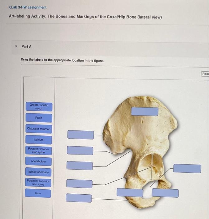
Solved Lab 3 Hw Assignment Art Labeling Activity Regions Chegg Com
0 komentar
Posting Komentar